
BHMS 1st year · Anatomy, Histology & Embryology
Abdomen, Pelvis and Perineum — the first-year Anatomy chapter below the diaphragm
A study guide to Abdomen, Pelvis and Perineum in first-year Anatomy: thirty-seven lines sorted into seven groups you can plan around.
Vaibhav Kumar Rai · · 4 min read
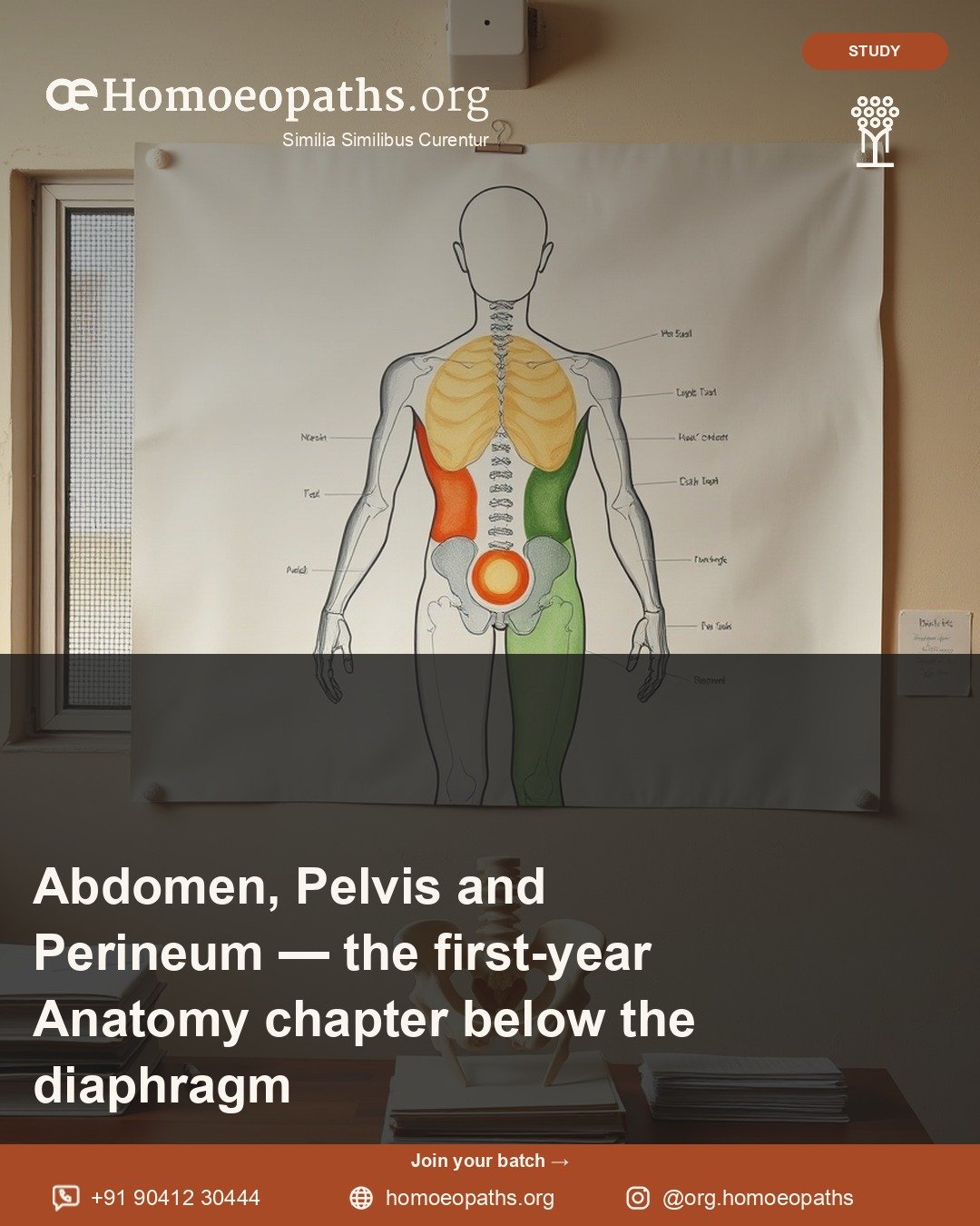
This chapter is big. The sheet gives it thirty-seven lines. That is more than any other chapter in first-year Anatomy. So the first job is not to memorise. The first job is to see the shape.
The NCH syllabus lists “Abdomen, Pelvis & Perineum” as chapter seven of Anatomy, Histology and Embryology (HomUG-AHE). It covers everything below the diaphragm: the belly, the bony basin under it, and the area at the very bottom of the trunk. This piece sorts the thirty-five topic lines between “Introduction” and “Revision” into seven groups.
Three words first
Abdomen is the belly, the space between the diaphragm and the pelvis. Pelvis is the basin-shaped region below it, ringed by bone. Perineum is the region at the lowest part of the trunk, below the pelvic floor.
Group one: the walls and the lining
- Anterior abdominal wall
- Posterior abdominal wall
- Peritoneum
The abdomen has a front wall and a back wall. The peritoneum is a thin, smooth lining. It covers the inside of the walls and wraps many of the organs. Learn the peritoneum early. It helps you understand where every other organ sits.
Group two: the digestive organs
- Stomach
- Duodenum
- Jejunum and ileum, superior mesenteric artery
- Caecum and appendix
- Large intestine
- Rectum
- Anal canal
Read this list in order and you are following the path food takes. The duodenum, jejunum and ileum are the three parts of the small intestine. The caecum is where the large intestine begins, and the appendix hangs from it. The sheet pairs the jejunum and ileum with the superior mesenteric artery, the vessel that supplies much of the gut.
Group three: the glands and organs beside the gut
- Liver
- Gall bladder and extrahepatic biliary apparatus
- Pancreas
- Spleen
“Extrahepatic” means outside the liver. The biliary apparatus is the set of ducts that carry bile. So this line asks for the gall bladder and the bile ducts that lie outside the liver.
Group four: the blood vessels
- Abdominal aorta
- Portal venous system
The abdominal aorta is the main artery running down the back of the abdomen. The portal venous system is a special set of veins. It carries blood from the gut to the liver before that blood goes back to the heart. Draw it once, clearly. It links the gut, the liver and the heart in one picture.
Group five: the urinary organs and the suprarenals
- Kidney
- Supra renal glands
- Ureter
- Urinary bladder
The kidneys make urine. The ureters carry it down. The bladder stores it. The suprarenal glands sit on top of the kidneys, which is where their name comes from.
Group six: the reproductive organs
- Ovary, uterus, fallopian tube
- Prostate gland
- Scrotum and testis, vas deferens
The female and male organs are both on the sheet. For each, learn position, parts, blood supply and relations to nearby organs. The vas deferens is the duct that carries sperm from the testis.
Group seven: the pelvic walls and the perineum
- Walls of pelvis including pelvic diaphragm
- Perineum: superficial and deep perineal pouches
- Ischiorectal fossa
The pelvic diaphragm is the muscular floor of the pelvis. The perineal pouches are two spaces, one superficial and one deep. The ischiorectal fossa is a space beside the anal canal. These three topics are about spaces and layers. Diagrams will help more than long notes.
The embryology and histology lines
The chapter also carries six lines from other parts of the subject.
- Systemic embryology: development of the digestive system, and of the urogenital organs.
- Systemic histology: the digestive system, the urinary system and suprarenal gland, the male reproductive system, and the female reproductive system.
“Urogenital” means the urinary and reproductive organs together. These six lines build on chapter two, Embryology, and chapter three, general Histology. Give them their own place in your plan.
How the chapter sits in the year
It follows Thorax, across the diaphragm. It comes before Head, Neck and Face. First-year Physiology returns to many of these organs in its Digestive System, Renal Physiology and Reproductive System chapters. Study the structure here, and those chapters will make more sense.
What to take from it
- See the chapter as seven groups before you start any one topic.
- Learn the peritoneum early. It explains where the organs lie.
- Draw the portal venous system as one clear picture.
- Plan time for the embryology and histology lines from day one.
The sheet names topics. It does not set depth. Your textbook, the dissection hall and your teachers do that.
Where to go next on the platform
This piece sits in the first-year Anatomy, Histology and Embryology room. “Thorax — the first-year Anatomy chapter on the chest, the heart and the lungs” is the chapter before it. “Histology (General) — the first-year Anatomy chapter where the body goes under the microscope” lays the ground for this chapter’s four histology lines.
Written by Vaibhav Kumar Rai · Published by Homoeopaths.org · 21 October 2026
"Abdomen, Pelvis and Perineum — the first-year Anatomy chapter below the diaphragm." Homoeopaths.org. Published 21 October 2026. https://homoeopaths.org/learn/courses/bhms-1st-year/anatomy-histology-embryology/abdomen-pelvis-perineum-1st-year