
BHMS 1st year · Anatomy, Histology & Embryology
Central Nervous System: Brain — the first-year Anatomy chapter that closes the subject
A study guide to the Brain chapter of first-year Anatomy: coverings and fluid, spinal cord, brainstem, cerebellum, cerebrum, blood supply and cranial nerves.
Vaibhav Kumar Rai · · 4 min read
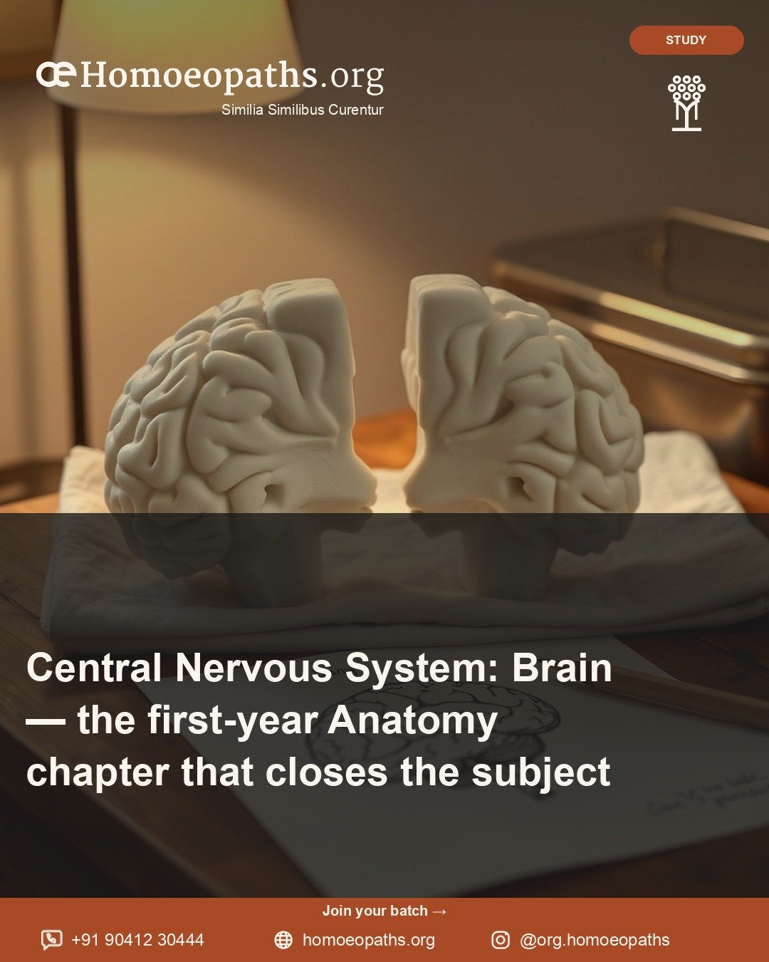
The brain is the last chapter of first-year Anatomy, and it can be hard to picture. The parts are deep, curved and folded around each other. You cannot see most of them from the outside. So this chapter asks for a different skill: building a picture in your head, layer by layer.
The NCH syllabus lists “Central Nervous System: Brain” as chapter nine of Anatomy, Histology and Embryology (HomUG-AHE). The sheet has nineteen lines, with “Introduction” first and “Revision” last. Between them are seventeen topics. They can be read from the outside in, and from the bottom up.
Start from the outside: coverings and fluid
Meninges and CSF. The meninges are the three coverings of the brain and spinal cord. CSF stands for cerebrospinal fluid. It is the clear fluid that bathes the brain and cord.
Blood supply of brain. The arteries that feed the brain and the veins that drain it. The head and neck chapter already brought these vessels up to the skull. Here they enter.
Then go from the bottom up
The central nervous system is the brain and the spinal cord together. The sheet climbs it in order, like floors of a building.
Spinal cord. The long cord inside the backbone.
Medulla oblongata, pons and mid-brain. Three parts that sit one above the other, just above the spinal cord. Together they are often called the brainstem. Learn them as one stack, each with its own features.
Cerebellum. The “little brain” at the back, behind the brainstem.
Diencephalon: thalamus and hypothalamus. The diencephalon sits above the brainstem, deep in the middle of the brain. The thalamus and hypothalamus are its two parts on the sheet.
Cerebrum: external features. The large upper part of the brain, with its folds and grooves, seen from the outside.
Functional areas of cerebral cortex. The cortex is the thin outer layer of the cerebrum. Different areas of it do different jobs. This topic asks you to know where they are.
Basal ganglia. Groups of nerve cells deep inside the cerebrum.
White matter of cerebrum: corpus callosum and internal capsule. White matter is made of nerve fibres that link one area to another. The corpus callosum joins the two halves of the brain. The internal capsule is a tightly packed band of fibres deep inside.
The spaces inside: the ventricles
- Fourth ventricle
- Third ventricle
- Lateral ventricle
The ventricles are connected spaces inside the brain. CSF fills them and flows through them. The sheet lists three: the fourth, the third and the lateral ventricles.
A good study method: draw all the ventricles on one page, joined as one system. Then mark where each one sits beside the brain parts you just learned. The fourth ventricle sits near the pons, medulla and cerebellum, and the sheet lists it right after them. The third ventricle sits in the diencephalon. The lateral ventricles are in the cerebrum.
The nerves that leave the brain
Cranial nerves. Nerves that come straight from the brain rather than from the spinal cord. Your textbook will number and name them. For each one, use the same headings: where it leaves the brain, where it goes and what it does.
The embryology line
Systemic embryology: development of brain. How the brain forms before birth. It builds on chapter two, Embryology. A simple flow diagram, from early stages to the adult parts, is a good way to learn it.
How the chapter sits in the year
This is the final chapter of the subject. It builds on almost everything before it. Chapter one taught nerves in general. Chapter three showed nervous tissue under the microscope. Head, Neck and Face brought the vessels and the cranial cavity to the door of the brain.
First-year Physiology has its own Central Nervous System chapter. It studies neurons, synapses, reflexes, the thalamus, the hypothalamus, the cerebellum, sleep and more. Anatomy tells you where each part is. Physiology tells you what it does. Study them side by side if you can.
What to take from it
- Learn the brain from the outside in, then from the bottom up.
- Treat the medulla, pons and mid-brain as one stack.
- Draw the ventricles as one connected system.
- Use fixed headings for each cranial nerve.
- Keep the embryology line on your plan.
The sheet names the topics and nothing more. How deep each goes is for your textbook and your teachers.
Where to go next on the platform
This piece lives in the first-year Anatomy, Histology and Embryology room. “Head, Neck and Face — the first-year Anatomy chapter where many small parts share a small space” is the chapter just before. “Physiology and Biochemistry in the First Year — how the body works” shows the Physiology side of the nervous system.
Written by Vaibhav Kumar Rai · Published by Homoeopaths.org · 22 October 2026
"Central Nervous System: Brain — the first-year Anatomy chapter that closes the subject." Homoeopaths.org. Published 22 October 2026. https://homoeopaths.org/learn/courses/bhms-1st-year/anatomy-histology-embryology/central-nervous-system-brain-1st-year
More in BHMS 1st year · Anatomy, Histology & Embryology
Anatomy, Histology & Embryology
Abdomen, Pelvis and Perineum — the first-year Anatomy chapter below the diaphragm
A study guide to Abdomen, Pelvis and Perineum in first-year Anatomy: thirty-seven lines sorted into seven groups you can plan around.
Vaibhav Kumar Rai · 21 October 2026 · 4 min read
Anatomy, Histology & Embryology
Anatomy, Histology and Embryology in the First Year — the body, region by region
What HomUG-AHE covers: general anatomy, embryology, histology, and the body region by region, from the upper limb to the brain.
Vaibhav Kumar Rai · 30 September 2026 · 4 min read